About
Our name describes our approach: artificial intelligence and materials science working together. We train machine-learning models on published data and on our own high-throughput experiments, use them to choose which alloys and ceramics to make, and then process, test and examine those materials in the laboratory. The results of those experiments become new training data.
The materials we care about are those for extreme environments, where very high temperatures, intense radiation and reactive atmospheres act together: refractory high-entropy alloys, ultra-high-temperature ceramics, and plasma-facing and structural materials for fusion and fission energy.
With support from the NSF Energy Storage Engine in Upstate New York, we also apply the same data-driven approach to batteries: forecasting the health and remaining life of lithium-ion cells from their cycling data, and training students in battery data science.
 Artificial intelligence for materials discovery
 Artificial intelligence for batteries
 Materials under irradiation and plasma
 Oxidation and corrosion at extreme temperatures
 Multi-scale electron microscopy
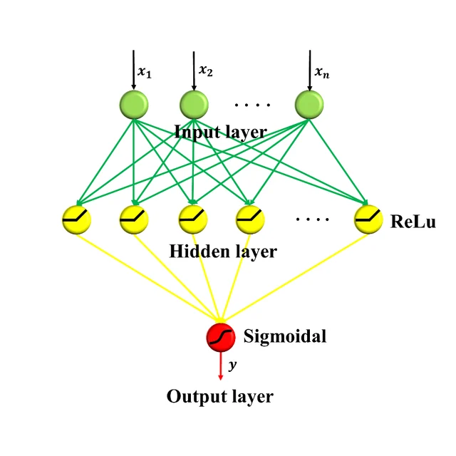
Artificial intelligence for materials discovery
High-entropy materials mix four, five or more principal elements, which opens a composition space far too large to explore one sample at a time. We train machine-learning models on curated literature data and on our own high-throughput experiments, use them to rank unexplored compositions, and then make and test the most promising candidates.
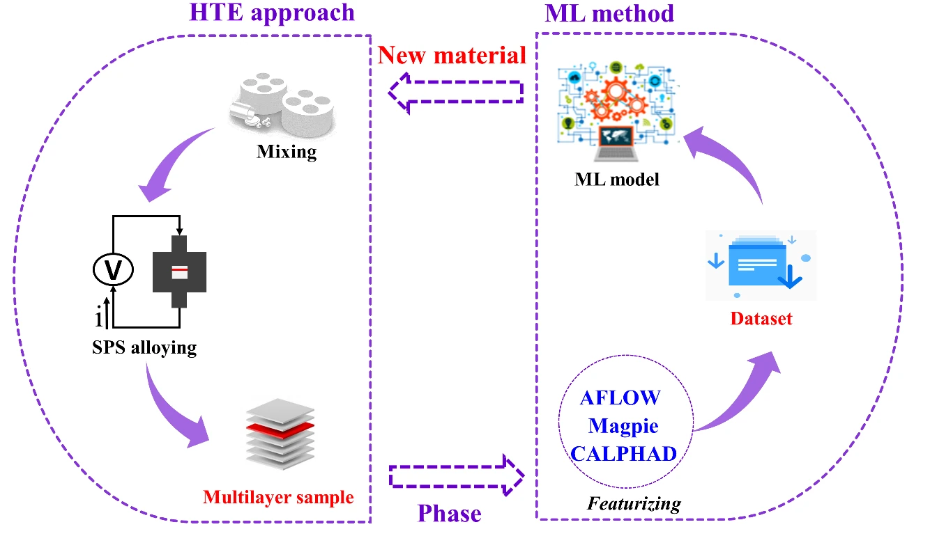
- Predicting single-phase refractory high-entropy alloys. Classifiers trained on a dataset of about 1,800 alloys identify compositions likely to form a single solid solution; ten of the predicted alloys were synthesized, and all formed a single BCC phase. Comput. Mater. Sci. 199 (2021) 110723
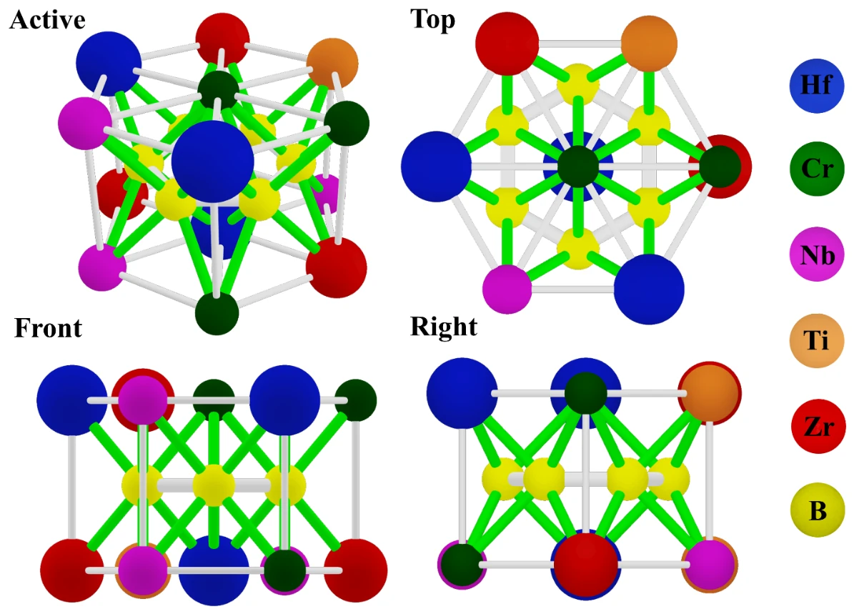
- A formation rule for high-entropy diborides. High-throughput experiments combined with interpretable machine learning showed that the mismatch in boron–metal bond lengths among the constituent diborides governs whether a high-entropy diboride forms a single phase, which gives a simple rule for predicting it. Acta Mater. 253 (2023) 118955
- Searching for oxidation-resistant ultra-high-temperature ceramics. Machine-learning models trained on high-throughput oxidation data for diboride–SiC composites pointed to new compositions, five of which were made and tested. Corros. Sci. 223 (2023) 111457
- Strength from composition. Machine-learning models estimate the intrinsic strength of refractory high-entropy alloys from composition-based features. Tungsten 5 (2023) 531–538
- Taking stock of the field. A review of machine learning for phase prediction in high-entropy alloys, and a perspective on chemical short-range order as an alloy-design tool. Tungsten 5 (2023) 32–49 · Curr. Opin. Solid State Mater. Sci. 41 (2026) 101254
Selected papers
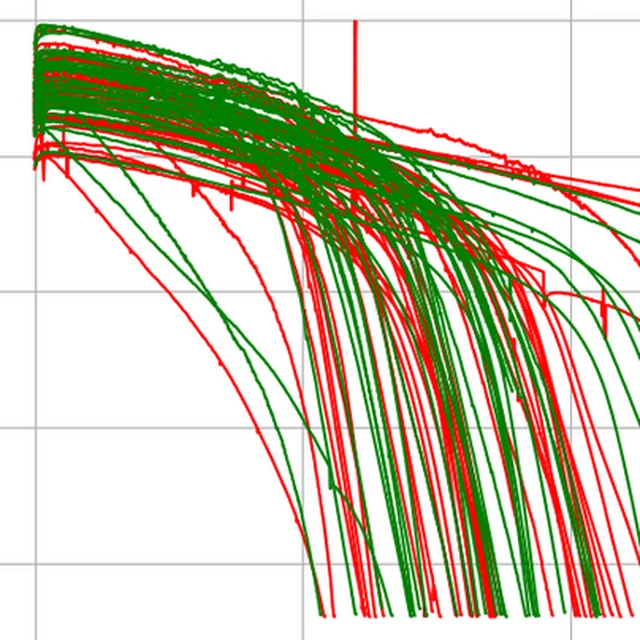
Artificial intelligence for batteries
A lithium-ion cell loses a little capacity with every charge and discharge, and no two cells age in quite the same way. Knowing a cell's state of health (SOH) and its remaining useful life (RUL) ahead of time matters for electric vehicles, for grid storage and for giving used batteries a second life. As part of the NSF Energy Storage Engine in Upstate New York, we build machine-learning tools that predict both from battery cycling data, and we use the same projects to train students in battery AI.
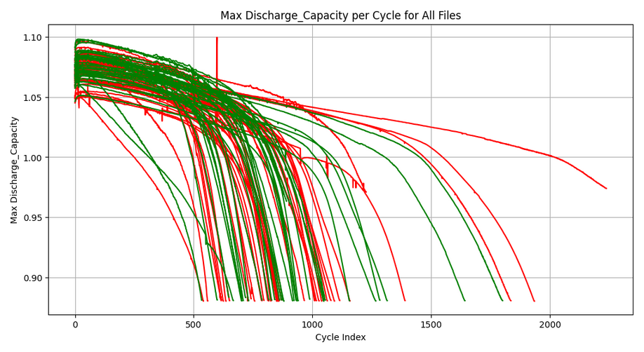
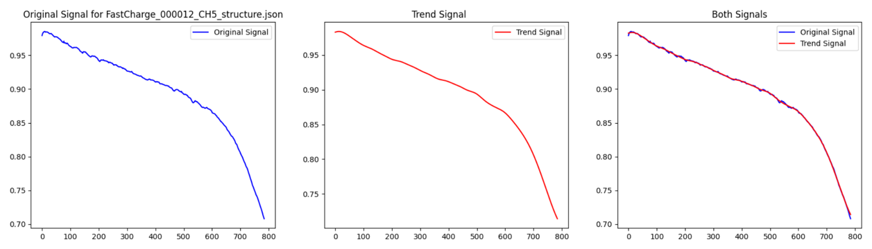
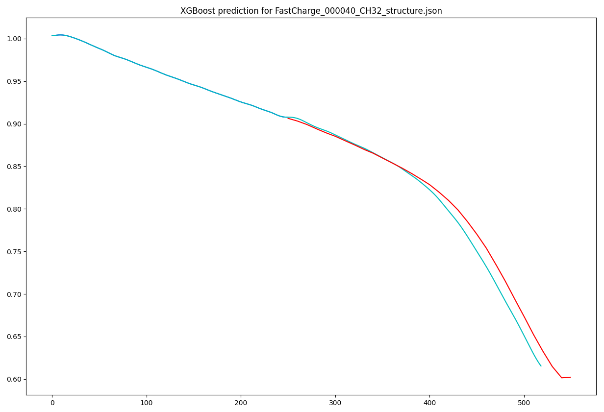
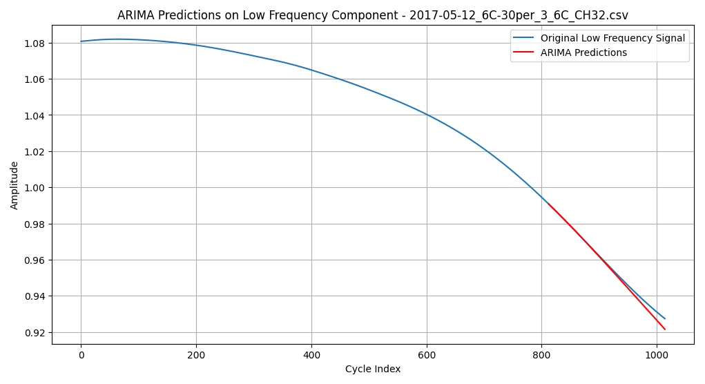
- Forecasting capacity fade. A cell's capacity curve is first separated into a smooth trend and noisy fluctuations by signal decomposition (CEEMDAN). Models then extend the curve step by step toward the end of the cell's life: support-vector regression, LSTM networks and ARIMA in our first studies, and more recently gradient-boosted trees (XGBoost) working on a rolling window of earlier cycles, with additional input features computed with the physics-based battery modeling library PyBaMM. Summer research posters, 2025 and 2026
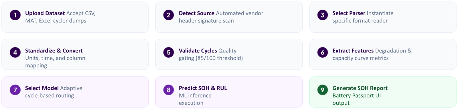
- A multi-source Battery Intelligence Platform. Battery test data come in many formats. This software reads cycling files in the formats of several public datasets and of a commercial battery cycler (NASA, CALCE, Oxford, Stanford and Arbin), converts them to one standard form, checks the quality of the data, and selects a model suited to the cell's cycle-life profile before reporting state of health and remaining useful life. Of the three tree-ensemble methods compared on held-out test data, Extra Trees performed best (R2 = 0.91 for SOH and 0.88 for RUL). Summer research poster and presentation, 2026
- Mining papers and databases for battery materials. Two exploratory projects looked at the materials side. One used a large language model and automated graph digitization to gather electrode, electrolyte and cycle-life data from more than 4,000 published papers into a single dataset for cycle-life prediction. The other built a physics-informed neural network from about 23,800 lithium transition-metal oxide entries in the Materials Project to screen cathode compositions by estimated volumetric energy density. Summer research posters, 2025
These projects are carried out by undergraduate researchers in the lab. The full list is under Battery AI workforce training.
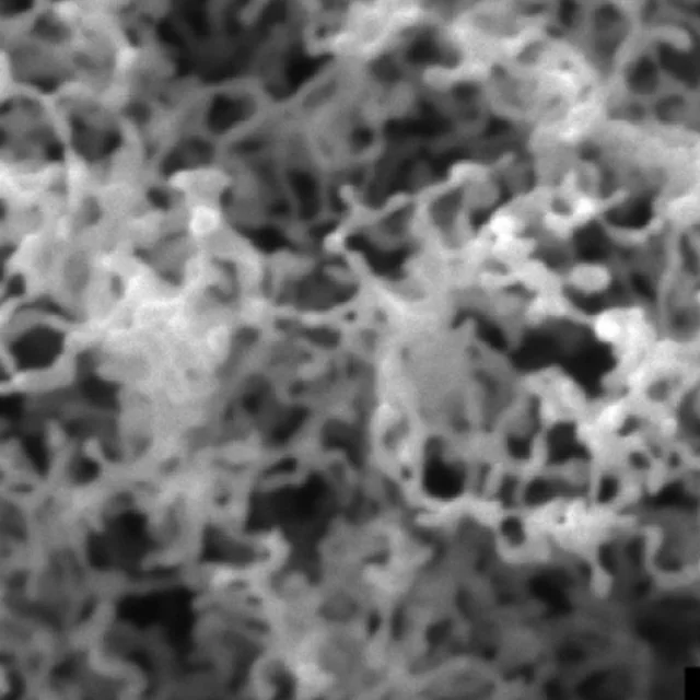
Materials under irradiation and plasma
Components that face a fusion plasma or sit in a reactor core are bombarded by helium, hydrogen isotopes and neutrons while running hot. We study how that exposure rearranges a material's microstructure, and which alloy designs resist it.
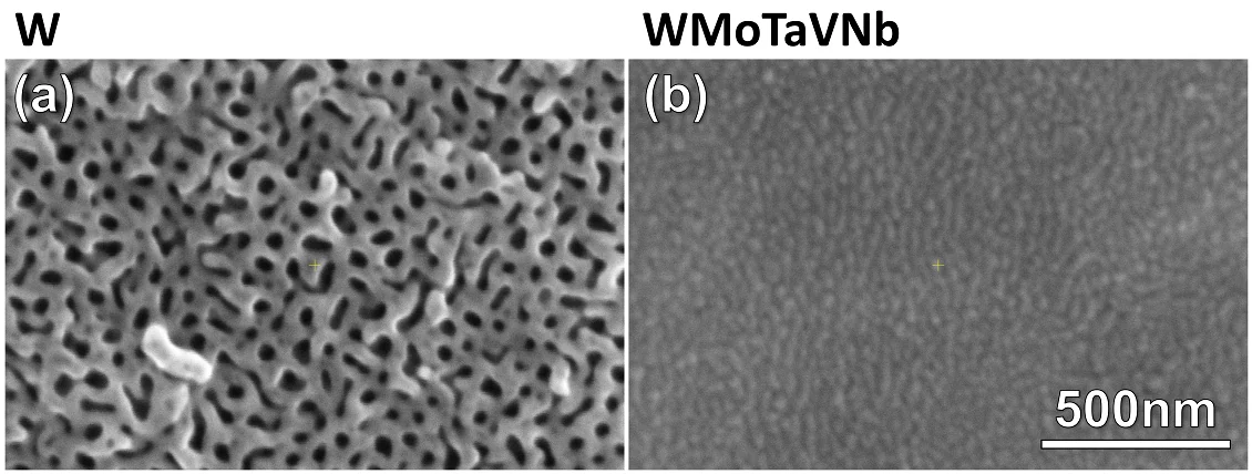
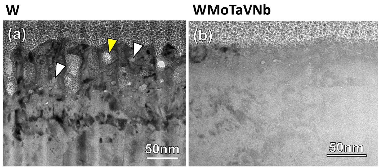
- Radiation-tolerant refractory high-entropy alloys. After the same helium plasma exposure, a tungsten-based high-entropy alloy (WMoTaVNb) grows far less surface “fuzz” than pure tungsten, and helium bubbles grow more slowly in it under helium-ion implantation. Molecular-dynamics simulations with collaborators link this to the sluggish motion of defects in the chemically complex alloy. J. Nucl. Mater. 588 (2024) 154761 · 595 (2024) 155060
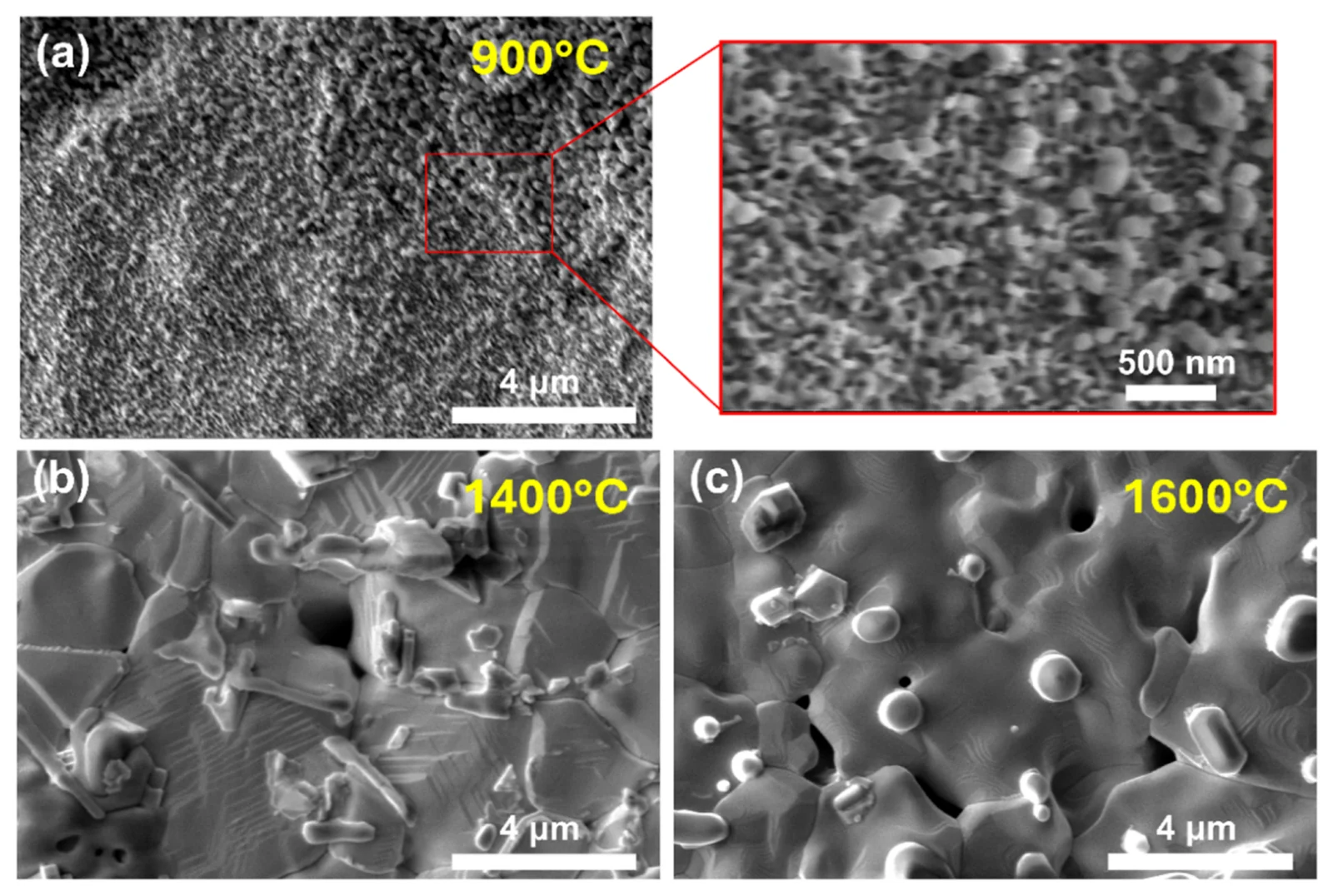
- How tungsten “fuzz” forms, grows and anneals. Electron microscopy of helium-plasma-exposed tungsten shows how nanotendrils nucleate, how the starting microstructure and the plasma flux change them, and how annealing above 900 °C restructures the fuzz back into the surface as its helium is released. Sci. Rep. 7 (2017) 42315 · Acta Mater. 124 (2017) 556–567 · J. Nucl. Mater. 509 & 510 (2018) · 628 (2026) 156641
- Irradiated tungsten and steels. In neutron-irradiated tungsten, transmutation products precipitate as second phases; in the reduced-activation steel Eurofer97, neutron irradiation drives segregation and phase instability; and in ferritic/martensitic steels irradiated with protons and neutrons, deformation twinning appears at high dose. Acta Mater. 165 (2019) 51–61 · J. Nucl. Mater. 547 (2021) 152834 · 501 (2018) 336–346
- Nanostructured alloys under heavy ions. With collaborators, we examined the microstructure and micromechanical response of a bulk nanostructured high-entropy alloy after heavy-ion irradiation at 500 °C. Scr. Mater. 235 (2023) 115628
Selected papers
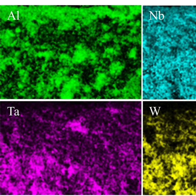
Oxidation and corrosion at extreme temperatures
Refractory alloys and ultra-high-temperature ceramics keep their strength where conventional alloys soften, but they must also survive oxygen. We combine oxidation and corrosion testing with microscopy from the micrometer to the atomic scale to find out what makes an oxide scale protective, and we use the measurements to train models that search for better compositions.
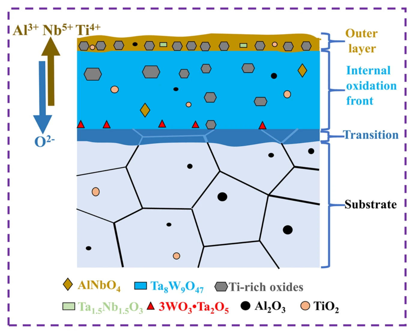
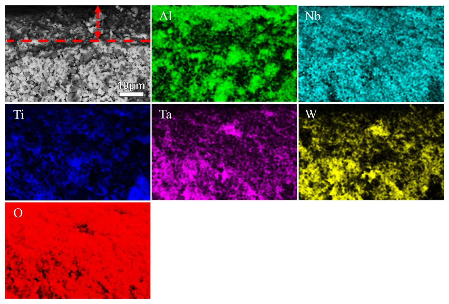
- Why WTaNbTiAl resists oxidation. Microscopy across length scales supports a diffusion-controlled, three-layer oxide scale model for the alloy's oxidation in air at 1000 °C. Corros. Sci. 204 (2022) 110377
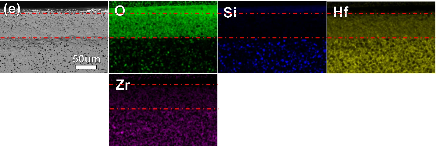
- Ultra-high-temperature ceramics at 1700 °C. Multicomponent (Hf,Zr)B2–SiC and (Hf,Zr,Cr)B2–SiC composites show oxidation resistance comparable to HfB2–SiC at lower density. Multi-scale microscopy traces this to a dense, multilayered oxide scale that is also smoother and less cracked, and should therefore be less prone to spallation. J. Mater. Res. (2026), doi:10.1557/s43578-026-01988-2
- Data-guided composition search. Oxidation measurements on multilayer samples train models that point to more oxidation-resistant diboride–SiC composites. Corros. Sci. 223 (2023) 111457
- Alloying for corrosion resistance. With collaborators, we studied how aluminum additions change the mechanical and corrosion behavior of a NbTaTiV high-entropy alloy. J. Alloys Compd. 1039 (2025) 183156
Selected papers
Multi-scale electron microscopy
Almost every question we ask ends at the microscope. We prepare site-specific specimens with a focused ion beam and examine them by scanning and transmission electron microscopy, diffraction and X-ray spectroscopy to link structure and chemistry to behavior.
- Site-specific specimen preparation. Focused-ion-beam cross-sections and TEM lamellae taken from oxide scales, irradiated surfaces and bulk ceramics.
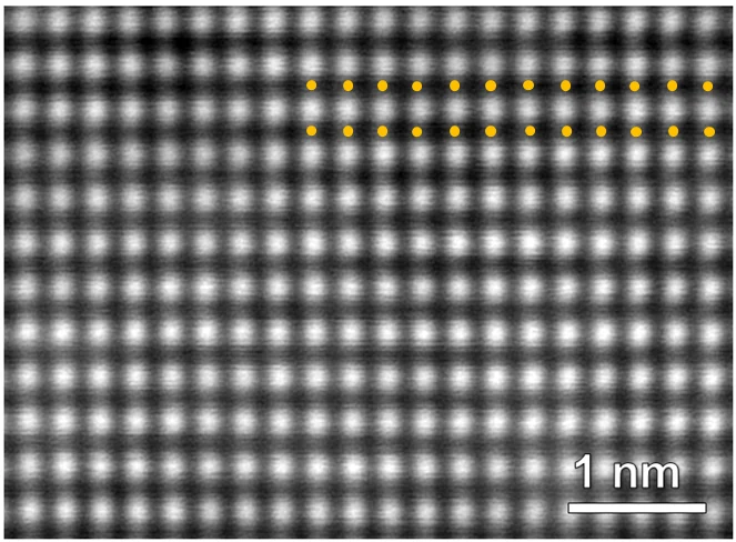
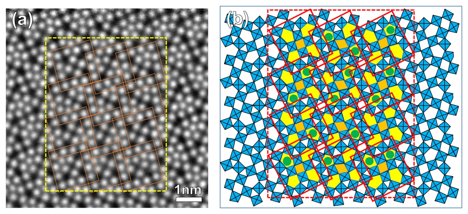
- S/TEM imaging, diffraction and spectroscopy. Bright- and dark-field imaging, electron diffraction, atomic-resolution STEM and EDS element mapping.
- Transmission Kikuchi diffraction. Orientation mapping of nanoscale grains, combined with STEM X-ray analysis for irradiated materials. Scr. Mater. 127 (2017) 132–135 · Microsc. Microanal. 23 (S1) (2017) 2218–2219
- Collaborative studies. We also contribute to joint work on high-entropy oxides and metal–organic frameworks, gradient-refractive-index chalcogenide glass-ceramics, borosilicate glasses and SiAlON ceramics. J. Am. Chem. Soc. 147 (2025) · Adv. Mater. 38 (2026) · J. Mater. Res. (2026) · J. Phys. Chem. C 130 (2026) · Int. J. Ceram. Eng. Sci. 6 (2024)
Facilities
The lab is based in Binns-Merrill Hall at Alfred University and makes heavy use of the university's electron microscopes.
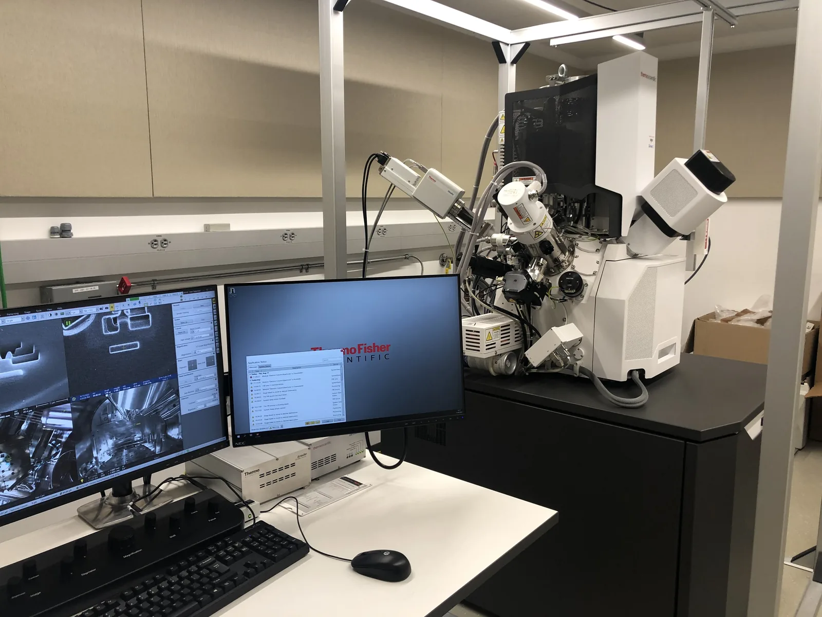 Focused ion beam – SEM
Thermo Scientific Scios 2 DualBeam with EDS, for site-specific cross-sections and TEM lamella preparation; acquired through NSF Major Research Instrumentation award 2018306 (co-PI: K. Wang).
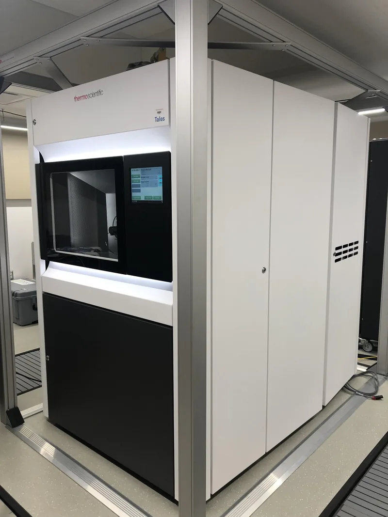 Transmission electron microscope
Thermo Scientific Talos F200X S/TEM (200 kV) with four in-column Super-X EDS detectors, for imaging, diffraction and element mapping, together with an in-situ heating and gas-environment holder.
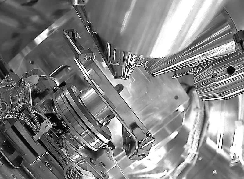 Sample preparation
Inside the FIB-SEM chamber, where cross-sections and TEM lamellae are cut from oxidized, irradiated and as-processed samples. Heat treatment and thermal aging are done in furnaces in Binns-Merrill Hall 127–130.
Support
Our recent work has been supported by faculty startup funds from the Inamori School of Engineering at Alfred University and by the U.S. Army (cooperative agreement W911NF-22-2-0061). Our battery AI research and workforce training are supported by the U.S. National Science Foundation through the NSF Energy Storage Engine in Upstate New York. The focused ion beam scanning electron microscope was acquired with support from the National Science Foundation (Major Research Instrumentation award 2018306).